Antioxidant Defense Mechanisms
Molecular Protection, Redox Homeostasis and Cellular Resistance to Oxidative Stress
Antioxidant defense mechanisms represent an essential component of cellular protection and biological homeostasis. These interconnected systems regulate the production, transformation and elimination of reactive oxygen species and other reactive molecules generated during normal metabolic activity and environmental exposure.
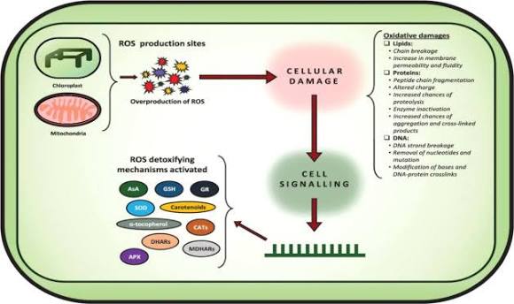
Under physiological conditions, reactive oxygen species participate in several important biological processes, including intracellular signaling, immune regulation, cellular adaptation and metabolic communication. However, excessive accumulation of these molecules can disturb redox balance and contribute to oxidative damage affecting proteins, lipids, nucleic acids and cellular membranes.
Antioxidant defense systems maintain an appropriate balance between oxidant production and antioxidant capacity through coordinated enzymatic and non-enzymatic mechanisms.
Their activity is particularly important in metabolically active tissues such as the nervous system, where neurons depend on mitochondrial energy production and precise regulation of intracellular signaling.
Understanding these protective mechanisms provides valuable insights into cellular resilience, oxidative stress regulation, mitochondrial function and the molecular processes involved in neuronal maintenance.
Understanding Oxidative Stress and Redox Homeostasis
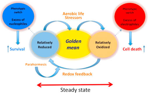
Oxidative stress occurs when the generation of reactive oxygen species and related oxidants exceeds the capacity of cellular antioxidant systems to maintain redox equilibrium.
Reactive oxygen species include several chemically distinct molecules, such as superoxide anion, hydrogen peroxide and hydroxyl radicals.
These molecules differ in their chemical reactivity, biological properties and potential effects on cellular structures.
Superoxide anion is generated through several enzymatic and metabolic processes, particularly within mitochondria. It can subsequently undergo conversion into hydrogen peroxide through spontaneous reactions or enzymatic activity.
Hydrogen peroxide is relatively more stable and can participate in intracellular signaling. However, excessive accumulation may contribute to oxidative damage, particularly in the presence of transition metals that facilitate the formation of highly reactive radicals.
Maintaining redox homeostasis requires a coordinated interaction between antioxidant enzymes, small-molecule antioxidants and regulatory signaling pathways.
Sources of Reactive Oxygen Species
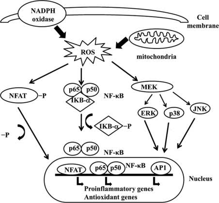
Reactive oxygen species originate from several intracellular sources.
Mitochondrial Electron Transport: During oxidative phosphorylation, electrons can escape from components of the respiratory chain and react with molecular oxygen, generating superoxide.
NADPH Oxidases: These enzymes produce reactive oxygen species through controlled electron transfer reactions and participate in cellular signaling and immune responses.
Peroxisomal Metabolism: Peroxisomes contain enzymes involved in lipid metabolism and other oxidative reactions that can generate hydrogen peroxide.
Xanthine Oxidase: This enzyme contributes to reactive oxygen species production during purine metabolism.
Inflammatory Responses: Activated immune cells can generate reactive oxygen species as part of their biological defense mechanisms.
The relative contribution of these sources depends on the cell type, metabolic conditions and physiological environment.

Enzymatic Antioxidant Defense Systems
Enzymatic antioxidants represent a primary line of defense against excessive reactive oxygen species accumulation.
These proteins catalyze specific reactions that transform reactive molecules into less harmful products.
The principal enzymatic antioxidant systems include superoxide dismutase, catalase and glutathione peroxidase.
Superoxide Dismutase
Superoxide dismutase is an essential antioxidant enzyme responsible for converting superoxide anion into hydrogen peroxide and molecular oxygen.
The reaction is represented by:
2 O₂•⁻ + 2 H⁺ → H₂O₂ + O₂
Several forms of superoxide dismutase are present in mammalian cells.
Cytosolic Superoxide Dismutase: This form contains copper and zinc as cofactors and is predominantly located in the cytoplasm.
Mitochondrial Superoxide Dismutase: This manganese-dependent enzyme is located in the mitochondrial matrix, where it contributes to the regulation of superoxide generated during cellular respiration.
Extracellular Superoxide Dismutase: This form is present in extracellular environments and contributes to the regulation of superoxide outside cells.
The activity of superoxide dismutase is closely connected to other antioxidant enzymes because its reaction produces hydrogen peroxide, which must subsequently be metabolized.
Catalase
Catalase is a heme-containing antioxidant enzyme that catalyzes the decomposition of hydrogen peroxide into water and oxygen.
The reaction is:
2 H₂O₂ → 2 H₂O + O₂
Catalase is particularly abundant in peroxisomes, although its distribution and activity can vary depending on the cellular environment.
By controlling hydrogen peroxide concentrations, catalase contributes to the maintenance of intracellular redox balance and limits the potential formation of highly reactive hydroxyl radicals.
Its activity is frequently evaluated in experimental investigations of oxidative stress and antioxidant protection.
Glutathione Peroxidase
Glutathione peroxidases represent a family of enzymes involved in the reduction of hydrogen peroxide and organic hydroperoxides.
Several members of this family contain selenium as an essential component of their catalytic structure.
These enzymes use reduced glutathione as an electron donor to convert peroxides into less reactive products.
The general reaction is:
H₂O₂ + 2 GSH → 2 H₂O + GSSG
In this reaction, reduced glutathione is oxidized into glutathione disulfide.
The regeneration of reduced glutathione is subsequently supported by glutathione reductase, which uses nicotinamide adenine dinucleotide phosphate in its reduced form.
This interconnected system contributes to the maintenance of cellular redox homeostasis.
Non-Enzymatic Antioxidant Defense Mechanisms
In addition to enzymatic protection, cells rely on non-enzymatic antioxidants that participate in the neutralization of reactive molecules and the regulation of oxidative processes.
These compounds may originate from endogenous metabolic pathways or external sources, including dietary components.
Their biological functions depend on their chemical properties, intracellular localization and interactions with other antioxidant systems.
Glutathione
Glutathione is one of the most abundant intracellular antioxidants.
It is a tripeptide composed of glutamate, cysteine and glycine.
Its sulfhydryl group allows it to participate in redox reactions and protect cellular components against oxidative modifications.
Glutathione exists mainly in two forms:
Reduced glutathione, which participates in antioxidant reactions.
Oxidized glutathione, which results from the oxidation of two glutathione molecules.
The ratio between reduced and oxidized glutathione is frequently used as an indicator of cellular redox status.
Maintaining an appropriate glutathione balance is particularly important for mitochondrial function, protein regulation and cellular survival.
Vitamin C
Vitamin C, also known as ascorbic acid, is a water-soluble antioxidant that participates in the neutralization of reactive oxygen species.
It contributes to the protection of aqueous cellular compartments and can interact with other antioxidant molecules.
Vitamin C also participates in several enzymatic reactions involved in collagen synthesis and other metabolic processes.
Its antioxidant activity is connected to the regeneration of certain oxidized antioxidant compounds.
Vitamin E
Vitamin E represents a group of lipid-soluble compounds, including tocopherols and tocotrienols.
Its principal biological function involves the protection of lipid membranes against oxidative damage.
Vitamin E can interrupt lipid peroxidation chain reactions by donating hydrogen atoms to lipid peroxyl radicals.
This mechanism helps limit the propagation of oxidative damage within cellular membranes.
Carotenoids and Other Dietary Antioxidants
Carotenoids are naturally occurring pigments found in numerous fruits and vegetables.
Some carotenoids, including beta-carotene and lycopene, have demonstrated antioxidant properties under specific biological conditions.
Other dietary compounds, such as polyphenols and flavonoids, have also been investigated for their potential interactions with oxidative and inflammatory signaling pathways.
Their effects depend on bioavailability, metabolism, concentration and the biological environment.
The Glutathione Redox Cycle and Cellular Protection
The glutathione system represents an important regulatory mechanism connecting antioxidant defense with cellular metabolism.
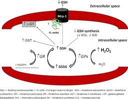
Reduced glutathione participates in the detoxification of hydrogen peroxide and organic hydroperoxides through glutathione peroxidase activity.
During this process, glutathione becomes oxidized into glutathione disulfide.
Glutathione reductase subsequently regenerates reduced glutathione using reducing equivalents supplied by NADPH.
This continuous recycling mechanism allows cells to maintain antioxidant capacity and regulate redox-dependent biological processes.
The availability of NADPH is therefore essential for maintaining glutathione-dependent antioxidant protection.
Several metabolic pathways contribute to NADPH production, including the pentose phosphate pathway.
The interaction between glucose metabolism, glutathione regeneration and antioxidant enzyme activity demonstrates the close relationship between cellular metabolism and redox regulation.
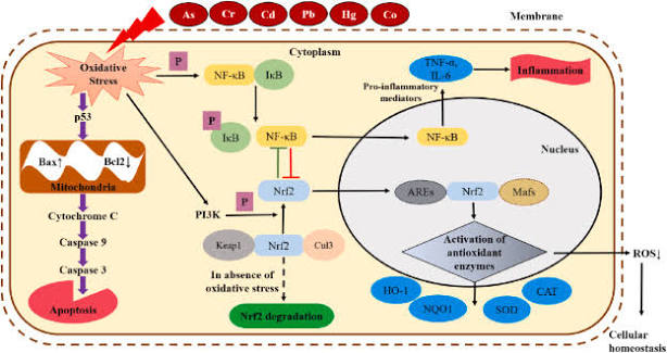
Nrf2-Mediated Regulation of Antioxidant Responses
The nuclear factor erythroid 2-related factor 2 signaling system is an important transcriptional regulator of cellular antioxidant responses.
It controls the expression of numerous genes involved in antioxidant defense, detoxification, metabolism and cellular protection.
Under unstimulated conditions, Nrf2 is regulated by Kelch-like ECH-associated protein 1, which facilitates its ubiquitination and proteasomal degradation.
Following oxidative or electrophilic stimulation, modifications in the regulatory system can reduce Nrf2 degradation and promote its accumulation.
Nrf2 subsequently translocates into the nucleus, where it interacts with small Maf proteins and recognizes antioxidant response elements within regulatory DNA regions.
This process promotes the expression of genes involved in cellular protection.
Important Nrf2-regulated proteins include:
Heme oxygenase-1.
NAD(P)H quinone dehydrogenase 1.
Glutamate-cysteine ligase.
Selected glutathione-related enzymes.
Several proteins involved in cellular detoxification.
The activation of this transcriptional system represents an important adaptive mechanism through which cells increase their antioxidant capacity.
Antioxidant Defense and Mitochondrial Function
Mitochondria are major regulators of cellular energy production and redox balance.
During oxidative phosphorylation, electrons move through the respiratory chain to support the production of ATP.
Although this process is essential for cellular metabolism, it can also generate reactive oxygen species.
Mitochondrial antioxidant mechanisms limit excessive accumulation of these molecules and help preserve mitochondrial integrity.
Manganese-dependent superoxide dismutase, glutathione peroxidases and peroxiredoxins contribute to mitochondrial redox regulation.
Mitochondrial quality-control mechanisms, including mitophagy, also participate in the removal of dysfunctional organelles.
These processes are particularly important in neurons because of their high energy requirements and limited capacity for cellular replacement.
Disturbances in mitochondrial antioxidant protection may influence membrane potential, ATP production, calcium regulation and cellular survival.
Antioxidant Protection in Neuronal Cells
Neurons are particularly sensitive to oxidative disturbances because of their metabolic activity, specialized membrane structures and dependence on mitochondrial function.
Their biological activity requires continuous energy production and tightly controlled intracellular signaling.
Oxidative stress may affect neuronal proteins, membrane lipids, mitochondrial components and nucleic acids.
Antioxidant defense systems therefore contribute to the preservation of neuronal integrity and functional communication.
Protection Against Lipid Peroxidation
Neuronal membranes contain substantial quantities of polyunsaturated fatty acids that are susceptible to oxidative modifications.
Lipid peroxidation involves a chain reaction initiated by reactive species that attack membrane lipids.
This process may alter membrane fluidity, receptor organization and cellular permeability.
Vitamin E, glutathione-dependent enzymes and other antioxidant mechanisms contribute to limiting lipid peroxidation.
Regulation of Neuronal Redox Signaling
Reactive oxygen species can function as signaling molecules when maintained within appropriate physiological ranges.
They participate in processes associated with synaptic activity, cellular adaptation and intracellular communication.
Antioxidant systems regulate these signals and prevent excessive oxidative accumulation.
The objective of cellular antioxidant regulation is therefore not the complete elimination of reactive oxygen species but the maintenance of an appropriate redox balance.
Neuronal Survival and Cellular Homeostasis
Antioxidant protection contributes to the regulation of mitochondrial function, protein stability and intracellular metabolic activity.
By limiting excessive oxidative damage, these systems support the maintenance of cellular structures and physiological processes.
Their interactions with inflammatory and survival-related pathways are particularly relevant to the investigation of neuronal stress responses.
Interactions Between Antioxidant Defense and Inflammatory Signaling
Oxidative stress and inflammation are interconnected processes that can influence one another through multiple molecular pathways.
Reactive oxygen species may modify signaling proteins and influence the activity of transcription factors involved in inflammatory regulation.
Similarly, inflammatory activation can increase the production of reactive molecules through mitochondrial activity and enzymatic systems.
Nuclear factor kappa B and Nrf2 represent two important regulatory systems involved in the relationship between inflammation and antioxidant protection.
Nuclear factor kappa B contributes to the regulation of inflammatory gene expression, while Nrf2 promotes the expression of several antioxidant and detoxification-related genes.
Their interactions are complex and depend on cellular conditions, signaling intensity and the nature of the stimulus.
Investigating these regulatory relationships helps explain how cells coordinate inflammatory responses with protective mechanisms.
Emerging Research Perspectives in Antioxidant Biology
Research into antioxidant defense mechanisms continues to evolve through the development of advanced molecular technologies and improved analytical approaches.
Current investigations increasingly focus on the integration of redox biology, mitochondrial regulation, transcriptional activity and cellular metabolism.
Important research directions include the characterization of Nrf2-dependent transcriptional responses, the investigation of mitochondrial antioxidant systems and the identification of molecular interactions between oxidative stress and inflammatory pathways.
Additional attention is directed toward the development of experimental approaches capable of distinguishing physiological redox signaling from excessive oxidative activity.
These developments contribute to a more detailed understanding of antioxidant regulation and its relevance to cellular adaptation.